Posted on

Treatments for Lazy Eye

Not for the Lazy, this takes a lot of work!

The treatment prescribed for you, or your child’s amblyopia will depend on various factors.

  • What type of amblyopia.
  • Age of the patient.
  • Compliance. 

Treatments include eye patches, drops, glasses or contact lenses, virtual reality headset, and sometimes surgery.

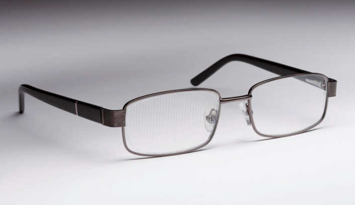
Corrective eyewear. Glasses or contact lenses can correct problems such as nearsightedness, farsightedness, or astigmatism that result in lazy eye.

Eye patches or occlusion therapy involves blocking the vision in one eye (generally the “good” eye) allowing the other eye to do all the work. (I told you this wasn’t for the lazy).

To stimulate the weaker eye, your child wears an eye patch over the eye with better vision for two to six or more hours a day. In rare cases, wearing an eye patch too long can cause amblyopia to develop in the patched eye. However, it’s usually reversible.

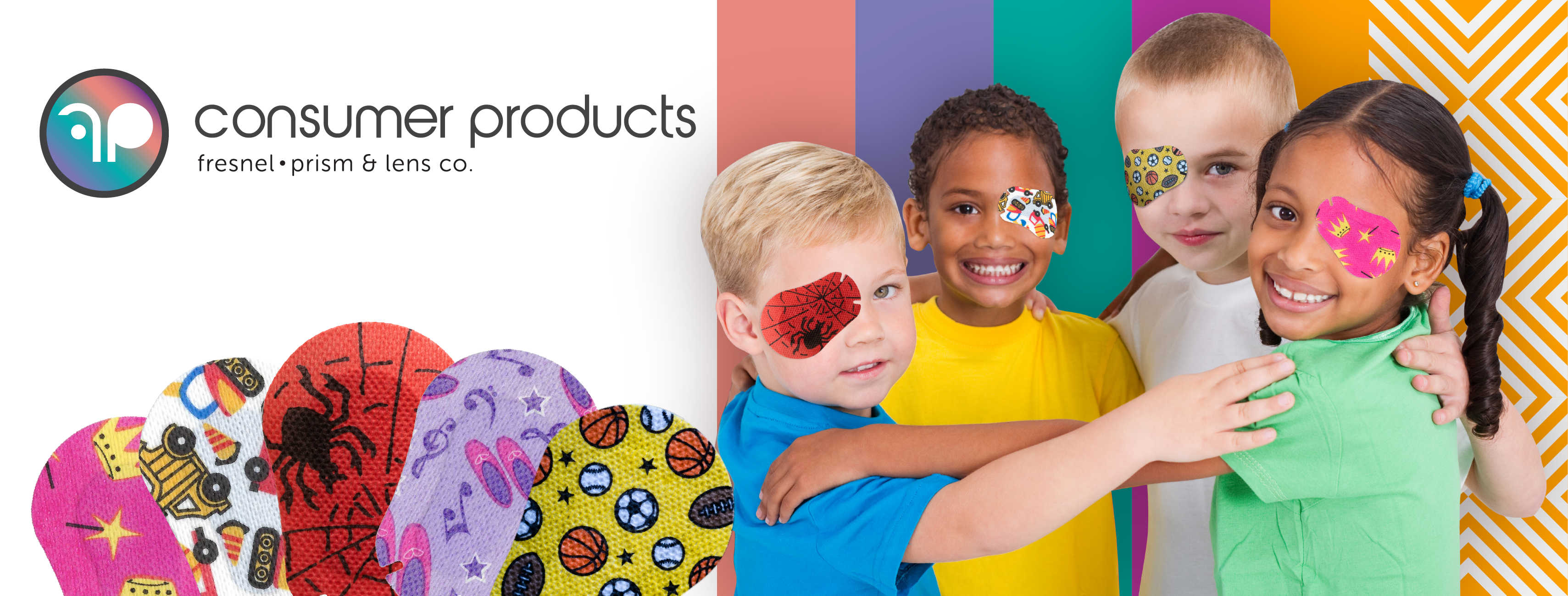
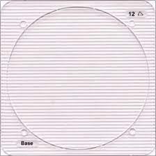
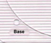
Eye patches are available as adhesive patches that adhere to the area around the eye, preventing any light or vison in the covered eye.  These may cause irritation to the sensitive skin around the eye, but there are tricks to help with this.  The majority of pediatric ophthalmologists will prescribe occlusion therapy with adhesive patches to ensure compliance.

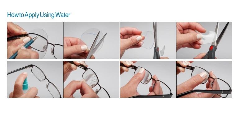
If a child wears glasses, a cloth eye patch may be an option. This type of patch attaches to the glasses to occlude the vision.  The drawback is the child has the opportunity to peek around or over the patch.  There is no skin irritation. They may be reused.

A pirate patch is a patch that has a strap that holds the patch over the eye.  The occlusion portion may be cloth, plastic, or silicone.  This is an option if glasses are not won.  This method is easy to peek around.

Eyedrops. An eyedrop of a medication called atropine (Isopto Atropine) can temporarily blur vision and is applied in the stronger eye. Usually prescribed for use on weekends or daily, use of the drops encourages your child to use the weaker eye and offers an alternative to a patch. Side effects include sensitivity to light and eye irritation.

Surgery. Your child might need surgery if he or she has droopy eyelids or cataracts that cause deprivation amblyopia. If your child’s eyes continue to cross or wander apart with the appropriate glasses, your doctor might recommend surgical repair to straighten the eyes, in addition to other lazy eye treatments.

Activity-based treatments — treatments offered may be drawing, doing puzzles, virtual reality headsets, or playing computer games. The effectiveness of adding these activities to other therapies may vary and has not been proved. However, research into new and non-traditional treatments is ongoing.

For most children with lazy eye, proper treatment improves vision within weeks to months. Treatment might last from six months to two years.

Remember, it is important for your child to be monitored for recurrence of lazy eye — which can happen in up to 25 percent of children with the condition. If lazy eye recurs, treatment will need to start again.

Lazy eye (amblyopia) – Diagnosis & treatment – Mayo Clinic

Occlusion Therapy – Eye Patches – Fresnel Prism and Lens (fresnel-prism.com)

Posted on

Which Eye Care Professional is for Me?

An eye care professional is an individual who provides a service related to the eyes or vision.

An ophthalmologist is a medical or osteopathic doctor who specializes in eye and vision care. Ophthalmologists differ from optometrists and opticians in their levels of training and in what they can diagnose and treat.

Ophthalmologists complete 12 to 14 years of training and education including medical school and are licensed to practice medicine and surgery. This advanced training allows ophthalmologists to diagnose and treat a wider range of conditions than optometrists and opticians. Typical training includes a four-year college degree followed by at least eight years of additional medical and surgical training.

An ophthalmologist diagnoses and treats all eye diseases, performs eye surgery, and prescribes and fits eyeglasses and contact lenses to correct vision problems. Many ophthalmologists are also involved in scientific research on the causes and cures for eye diseases and vision disorders. Because they are medical doctors, ophthalmologists can sometimes recognize other health problems that aren’t directly related to the eye, and refer those patients to the right medical doctors for treatment.

Specialties may include:

  • Pediatric Ophthalmology
  • Neuro Ophthalmology
  • Ocular Oncology

Optometrists are healthcare professionals who provide primary vision care ranging from vision testing and correction to the diagnosis, treatment, and management of vision changes. An optometrist is not a medical doctor. An optometrist receives a Doctor of Optometry (OD) degree after completing 2 to 4 years of college-level education, followed by four years of optometry school. They are licensed to practice optometry, which primarily involves performing eye exams and vision tests, prescribing, and dispensing corrective lenses, detecting certain eye abnormalities, and prescribing medications for certain eye diseases in some states. Many ophthalmologists and optometrists work together in the same offices, as a team.

Specialties may include:

  • Pediatric Optometry
  • Neuro Optometry
  • Low Vision Optometry

Opticians are technicians trained to design, verify, and fit eyeglass lenses, framescontact lenses, and other devices to correct eyesight. They use prescriptions supplied by ophthalmologists or optometrists, but do not test vision or write prescriptions for visual correction. Opticians are not permitted to diagnose or treat eye diseases.

Orthoptists are the experts in diagnosing and treating disorders in eye movement and problems with how the eyes work together; called binocular vision. These can be caused by issues with the muscles around the eyes or defects in the nerves enabling the brain to communicate with the eyes.

They are trained to offer a range of non-surgical treatments, including eye patches, eye exercises, prisms, or glasses. They also commonly work with patients dealing with neurological conditions, such as stroke, brain tumors, or multiple sclerosis.  Orthoptists are uniquely trained to treat disorders such as amblyopia, genetic disorders, and complex pediatric and adult strabismus.

Optometrist vs. Ophthalmologist: How to Choose an Eye Doctor (allaboutvision.com)

FAQs – Fresnel Prism and Lens (fresnel-prism.com)

Posted on

How the Eye Works

The Eye

Crash course on the EYE.

  • The eye is the only organ of the body that you can see into without having to cut it open or stick a tube into!
  • Approximately 80 percent of everything we learn comes through our eyes.
  • The eye contains over two million working parts and is considered the second most complex organ in the body— the most complex is the brain.
  • The optic nerve is critical to your vision. It’s an extension of your central nervous system, which includes your brain and spine. The optic nerve transmits electrical impulses from your eyes to your brain.

The eye works much like a camera.

  • Both have a lens or lens system.
  • Both provide a way to collect incoming light.
  • Both provide an image that must be interpreted by the brain.

Our brain works together with our eyes to process the information we see and transform it into recognizable images. The clear disc-like part of the eye called the lens helps to focus light on the retina. The ciliary muscle adjusts the shape of the lens, helping it to focus on objects at different distances. This automatic focusing is a reflex response and is not controlled by the brain.

Once the image is clearly focused on the sensitive part of the retina, the energy in the light that makes up that image creates an electrical signal. Nerve impulses can then carry information about that image to the brain through the optic nerve, which is a collection of more than a million nerve fibers. As the cornea bends light when it enters the eye, the brain receives images that are upside down, so it turns them the right way up when it processes the information.

We can “see”!  Yes, vision is a great deal more complex than this, but you get the point.

https://www.fresnel-prism.com/about-us/

Posted on

Lazy Eye – Amblyopia

Before we start, I just want to share some cool information. The eye is an extension of the brain, and contains nerve tissue in the light sensitive layer, the retina. Compared to the brain, the retina of the eye offers the distinct advantage of being accessible to high resolution imaging using advanced light and optical technology. Pretty Cool.

Amblyopia (also called lazy eye) is a type of decreased vision that usually happens in just one eye, but less commonly in both eyes. It develops when there’s a breakdown in how the brain and the eye work together, and the brain can’t recognize the sight from one eye. This affects between 2% -4% of children.

With amblyopia, the brain gets a blurry picture from one or both eyes and so it does not learn to see well. Amblyopia can often be fixed in childhood, but if not treated, it can cause permanent vision loss.  It is the main reason that kids lose vision. (AAPOS.org)

Amblyopia can be tricky for parents or caregivers to spot because children don’t usually complain about blurry vision. They will use the eye that sees better and won’t notice that anything is wrong. Also, the weaker eye may look normal.  Many times, a problem will only be found during a vision test. Vision Screening, recommended by the American Academy of Pediatrics (AAP), helps to catch amblyopia early when it can still be treated.
There are three defined types of Amblyopia:

Refractive amblyopia – This is the most common type. It happens when a child needs glasses.

Strabismic amblyopia– This is caused by eye movement problems. One eye may turn in, out, up, or down. When this happens, the brain begins to ignore, or “turns off” the wandering eye and does not learn to see well.

Deprivational amblyopia – Rarely, problems like ptosis (a droopy eyelid), cataract (cloudy spot in the lens/focusing part of the eye) or cornea scar (cloudy spot in the clear front part of the eye) can stop a child from seeing. If not treated very early, these children never learn to see very well and can have very poor vision.

The treatment depends on the type of amblyopia that the child has. 
Glasses help some children with amblyopia, especially those with refractive amblyopia and strabismus that gets better with glasses (such as accommodative esotropia). 
 
For many children, glasses may not be enough to help the amblyopia. Some children need treatment with eye patching or eye drops in the stronger eye to make it weak and to force the brain to use the weaker eye more. Children with a droopy eyelid, cataract, scar or eye movement problem may need surgery to help the amblyopia.

https://medriva.com/health/a-new-dawn-in-amblyopia-treatment-harnessing-technology-to-see-the-world-anew

Finally! Eye Patches.
This is where we come in!
We are the makers of the MYI Occlusion Eye Patch and the CoverClude Occlusion Eye Patch.
An eye patch is used to cover or occlude the “good” eye to make the “bad” eye work and
force the brain to use the weaker eye more to make the vision better. 

MYI Occlusion Eye Patches and the CoverClude Occlusion Eye Patch are adhesive patches that adhere to the skin around the eye to stop any “peeking. Cloth patches and pirate patches can be moved to allow the child to see around the patch – not good.

  • Our patches are made in America – Minnesota to be exact – very good.
  • Other patches are made in Europe or China. Please support small US business.
  • We developed the black occlusion layer to eliminate light reaching the eye. Cool.
  • Our adhesive is a medical grade adhesive, again American made. Wow!


You pick what designs you and your child want in your pack – this increases compliance when your child has an involvement in the decisions. – Power to the child.

3M Fresnel Optics – Fresnel Prism and Lens (fresnel-prism.com)

Lazy eye (amblyopia) – Symptoms & causes – Mayo Clinic

Youtube

Posted on

Patch Irritation and Removal

Suggestions to Help with Patch Irritation and Removal

Many kids experience a rash or irritation from the adhesive tapes used with patches. Here are a few ideas to try if you experience this.

  • Apply an adhesive barrier before placing the patch on. The patch adheres to the film and not the skin.
  • Before applying the patch, use either Vitamin E – pierce the capsule and use the gel – or a liquid anti-acid (Mylanta or Milk of Magnesia) and apply a thin coat around the eye. Let it dry, and then put on the patch.
  • Before applying the patch, please put it on your arm or pant leg to degrade the adhesive strength so it will be less sticky.
  • If certain areas have sores, cut the adhesive around these areas if possible so they don’t stick there, but do it everywhere else.
  • Alternate turning the patch small side in toward the nose, then the next day, large size toward the nose if this keeps the adhesive from the area with the sores.
  • When removing the patch, try coating the outside with Vaseline, baby oil, Aquaphor, or diaper ointment for about 15 minutes before removing it to loosen the adhesive.

Redness from eye patches – Fresnel Prism and Lens (fresnel-prism.com)

Posted on

HELP! My Child Will Not Keep the Patch On.

You are not alone.

Imagine if you have good vision in one eye but not so great vision in the other eye, and you have to cover the only eye you see well from. You would try to get that patch off, too.

But because you are not alone, we have collected tips and ideas that have worked for other families, and they may work for you too, at least for a bit; every extra minute is a win.

  • Special playtime toys are only available during patching.
  • One-on-one activities.
  • Helping to cook.
  • Bubbles.
  • Dance party.
  • Toddler age? Make patching time active learning time. This encourages both sides of the brain to interact and is better for this age than traditional eye exercises.
  • Movie time.
  • Apply the patch first thing in the morning, before they wake if possible, so it does not seem like you have taken something from them.
  • Make patching part of the daily routine.
  • Use a timer that will let them know when the time is completed. That way, you are not the bad guy saying it has not been long enough.
  • Rewards for short-term goals. 
  • Do not give time off from patching for good behavior, like if your child requires an insulin shot – they must do this to remain healthy. NO Option.

Innovative Solutions to Eye Occlusion Therapy – Fresnel Prism and Lens (fresnel-prism.com)

FAQs – Fresnel Prism and Lens (fresnel-prism.com)

Posted on

What about Low Vision Patients, TBI Patients, Stroke Patients?

3M Press-On™ Optics: Prisms and Plus Power Aspheric lenses are the tools needed to help the patient.

After a stroke, a patient may experience vision problems, which may improve over time. Instead of waiting for the vision to stabilize, using the 3M Press-On™ prism may allow the patient to have a quality of life doing familiar things while they recover. With the cost of a ground-in prism being prohibitive to change often as the patient recovers, using an array of Press-On™ prisms may expedite the recovery. Therapy will be more successful when the patient is able to engage.

With TBI patients, it has been questioned how a patient can re-learn particular skills if they are unable to see correctly. With resulting double vision or eye misalignment, a patient is not able to re-learn to walk safely or will experience severe headaches as the brain is trying to heal. Using a Press-On™ prism may assist when the headaches are due to the visual stress of the eye/brain attempting to correct the alignment.

Low vision is a vision problem that makes it hard to do everyday activities. It can’t be cured with glasses, contact lenses, or other standard treatments like medicine or surgery, but it can be treated to provide the best vision possible. Press-On™ Aspheric lenses may be used to magnify by placing on the lower portion of each lens. This is a lightweight solution that may allow the patient to manage day-to-day activities without the use of a high-plus reading loupe.

Discover the Power of 3M™ Press-On™ Optics – Fresnel Prism and Lens (fresnel-prism.com)

technical-information.pdf (3m.com)

Posted on

Why Prescribe a Press-On™?

A 3M Press-On™ is a simple, therapeutic, inexpensive way to correct various visual disorders.

  • Strabismus – early onset
  • Mild Amblyopia
  • Nystagmus
  • Inoperable Strabismus
  • Incomitant Strabismus
  • Diplopia
  • Phorias

A 3M Press-On™ provides immediate correction. Happy patient.

Press-On™ Optics adds no noticeable weight or thickness to the patient’s spectacle lens. 

The patient is looking good!

Improve the quality of life for your patient simply, inexpensively, and fast! Patient will love you!

Hint: It is best to maintain an inventory of a few of the prisms you use the most to provide corrective action and have the patient walk out of the office better than they walked in. We receive phone calls from patients asking if they can purchase a prism from us – the answer is yes; we want them to have the doctor who has prescribed the prism apply it correctly. 

We require they provide us with a copy of the current prescription before selling them a prism. Now, the problem arises as the patient (who has vision problems) is supposed to apply the prism correctly. These are frustrated and scared patients looking to their eye care provider for the solution to their problem. 

3M Fresnel Optics – Fresnel Prism and Lens (fresnel-prism.com)

why prescribe press on fresnel lens – Search Images (bing.com)

Posted on

The magic of a Fresnel lens

Ever see a lighthouse beam and wonder how it shines so brightly across vast distances? Or perhaps you’ve used a thin plastic magnifier to enlarge tiny text. You may thank a Fresnel prism lens for this magic.

A prism is a wedge-shaped piece of transparent material cut with precise angles and plane faces. The path of light entering through a prism will be affected / changed as it passes through the prism and then exits the prism. How much change will be dependent upon the prism material and the prism angles and the angle the light first enters the prism surface. Fresnel prisms are made from a thin transparent plastic sheet consisting of multiple angular grooves on one side. These prisms are much lighter than the regularly used prisms of the same power.  It can be thought of as a series of tiny plastic prisms placed on a plastic platform that is thin and adjacent to each other in a format that the base of one is attached to the apex of the next prism. The magnification effect seen with conventional prisms is minimal with Fresnel prisms.

After 1960, the Fresnel principle, which existed for more than 150 years, was used for the first time to manage strabismus. In 1970, the Optical Scientific Group from San Rafael, California designed a combination of a series of Fresnel press on prisms. These prisms are made up of plastic membranes with powers ranging from 0.5-30 prism diopters. The Fresnel prism is derived from the concept of hand-ground lenses prepared by the physics and French specialist Augustine Fresnel in 1921.

Initially, Fresnel prisms were intended for use in lighthouse beacons. Prisms have been used extensively in the field of ophthalmology, especially related to neuro-ophthalmology and strabismus. Prisms are used extensively to increase the binocular field, relieve diplopia, or expand the field of vision. Fresnel prisms are thin transparent plastic membranes with multiple tiny prisms attached from base to base. Augustin Fresnel articulated the optics of Fresnel prisms.

The angle of refraction of a prism depends upon the angle of the surface and the refractive index of the material of which the prism is made. The angle of refraction is independent of the thickness of the prism. A Fresnel prism offers the advantage of being lightweight and easy to handle. The most common use is to temporarily relieve the diplopia in cases like nerve palsies secondary to trauma, blowout fractures, decompensated phoria, divergence insufficiency, or convergence insufficiency.

Fresnel prisms are most commonly used as temporary prisms. Traditionally, prisms have also been used in patients with stroke and homonymous hemianopia. The aim is to shift the peripheral image towards the central retinal meridian.

How Does it Stay on the Lens?

The 3M Press-On™ prism is applied to the inside of the carrier lens with just water.

The 3M Press-On™ prism is attached to a border which identifies the base, if the border is discarded the base may identified easily by running a finger across the prisms, it will slide smoothly towards the base and jagged away from the base.

The smooth side of the prism is attached to the lens.

Fitting a 3M Press-On™ Prism

  • Holding the prism in place against the inside of the lens with the base oriented as required.
  • Draw on the rough side of the prism around the shape of the lens with a non-permanent marker. It is best if the lens can be removed from the from frame to ensure the prism alignment is correct.
  • Cut the prism using a small pair of scissors or Exacto knife.
  • Cut the prism a bit smaller than the lens so no portion will overlap the frame and be pulled off.
  • Ensure the carrier lens is clean a free of dust and oil. Do not use cleaner with alcohol as this will damage the prism. There are dish soaps that contain alcohol.

Applying a 3M Press-On™ Prism

  • Put a few drops of water on the inside of the lens where the Fresnel prism is to be placed. Push the smooth side of the Press-On™ prism down on the water.
  • Using your thumb squeeze out any excess water and air bubbles and gently pat dry.
    The prism may appear cloudy or hazy for the first 24 hours until all the water has dissipated – it may be easy to slide during this time, so care is encouraged.
  • After it has dried the prism will no longer move. To remove the prism for the carrier lens, use the edge of your fingernail to carefully pull away.

The Fresnel Press-On™ prism can be cleaned using a non-alcohol lens cleaner and a soft micro-fiber cloth. Pull the cloth in the direction of the prisms vs against.

What Powers are Available?

The 3M Press-On™ Prisms are available in 17 Diopter Powers: IMAGES MISSING, may be on site
I found a few I put in folder.

Fresnel Prism to the Rescue (reviewofoptometry.com)

Posted on

Who is The Fresnel Prism and Lens Company?

Who is The Fresnel Prism and Lens Company?

The Fresnel Prism and Lens Company has been around since 1987. The Press-On™ Prism, patented by Arthur Jampolsky, MD, has been a staple in the eyecare professional’s toolbox since 1971. The Press-On™ Optics is based on the 1822 Fresnel Principle of a Fresnel Prism, which you will see in a few of the remaining old lighthouses. Many have asked us about the name Fresnel . . . the S is silent. 😊  

The Fresnel Principal was developed by French engineer Augustin Fresnel in 1822. It states: A prism’s angle of refraction depends only upon the angle of the surfaces and the index of refraction of the material of which the prism is made. The angle of refraction is independent of the prism’s thickness.  
Our main business will always be the 3M Press-On™ Optics. As 3M’s largest US distributor of Press-On™ Optics, we will always have a warehouse full of prisms to be shipped to you the same day.

We will always answer the phone with a smile – we pride ourselves in our over-the-top customer service.

We are still a Mom-and-Pop operation, plus two wonderful folks who take the orders, pack them, and ship them. Folks think we are a large operation, but nope, we are just super-efficient!

3M Fresnel Optics – Fresnel Prism and Lens (fresnel-prism.com)